Markus Meyer
Dr. rer. nat. Markus Meyer

Former: Department of Chemistry and Pharmacy
Former: Institute of Physical Chemistry II (Fink Group)
Egerlandstr. 3
91058 Erlangen
Germany
- Phone number: +49 9131 85-27320
- Fax number: +49 9131 85-28867
- Email: markus.meyer@fau.de
- Website: https://www.chemistry.nat.fau.eu/fink-group/
Project description
A fundamental constraint which limits the collection of artifact-free data is beam damage caused by intense irradiation. Various damage mechanisms have been reviewed and proposed elsewhere [1,2]. A more sophisticated understanding of the mechanism can lead to improved experimental routines, in order to acquire data with better quality. This issue becomes even more important with the next synchrotron generation and free electron lasers being established around the world.
Within this project we address the challenge of dealing with irradiation sensitive soft matter like polymers or organic thin films. Using a new all but in-situ fashioned variation of the STXM probe method, new quantitative insights into the mechanism of molecular fragmentation is expected. In the present context, soft X-ray Scanning Transmission Microscopy (STXM) uses the chemical fingerprint of near-edge X-ray absorption fine structure (NEXAFS) to follow intramolecular processes. Investigations are therefore focused, but not limited to the understanding of beam damage mechanisms in polymers, the influence of secondary processes on sample degradation during irradiation and the influence of deposition on analysis and misleading argumentation in the present literature.
The project studies are extended to biocompatible polymer materials using TEM, STXM and SAXS studies. Addition of elastomers into a PLA-Matrix material offers significantly improved mechanical properties [3]. Applying in-situ SAXS measurements, the growth, and domain formation of partially crystalline elastomer units within the PLA-Matrix can be followed and analyzed.
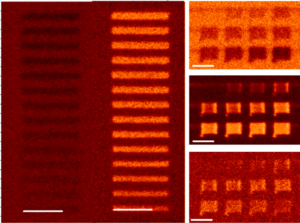
Figure 1: Systematic dose and energy-dependent beam damage studies on PMMA thin films
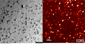
Figure 2: TEM and STXM image of elastomer inclusions in PLA matrix.
References
[1] R.F. Egerton et al., Micron 35 (2004) 399.
[2] A.F.G. Leontowich et al., J. Electron Spectrosc. Relat. Phenom. 206 (2016) 58.
[3] S. Cai et al., RSC Advances 6 (2016) 25531.