Andreas Hutzler
Dr.-Ing. Dr.-Ing. Andreas Hutzler
Department of Electrical, Electronic and Communication Engineering (EEI)
Chair of Electron Devices
Cauerstr. 6
91058 Erlangen
Germany
- Phone number: +49 9131 85-28638
- Fax number: +49 9131 85-28698
- Email: andreas.hutzler@fau.de
- Website: http://www.leb.eei.uni-erlangen.de/
Project description
Liquid cell transmission electron microscopy (LCTEM) is a novel characterization method that is already applied in a large diversity of scientific fields like biology [1], electrochemistry [2] and materials science [3]. It enables in situ investigations into dynamic systems in a fluid environment and thus real time observations of ongoing processes at the site of the event under realistic conditions. In order to achieve this, a thin layer of the specimen solution is enclosed between two electron transparent membranes and sealed to avoid vaporization inside the vacuum of the electron microscope.
Within the frame of this project, a new liquid cell design was developed [4,5] which is consisting of a robust silicon frame with a structured silicon nitride membrane. The membrane containing micrometer sized wells encapsulates the specimen with an additional graphene membrane that is placed on top (see Fig. 1). This advanced liquid cell design provides large viewing areas and angles. Therefore it is not only feasible for conventionally applied in situ TEM techniques like scanning TEM (STEM) or high resolution TEM (HRTEM) but offers improved performance in analytical methods like energy dispersive X-ray spectroscopy (EDXS) as well as electron tomography.
Issues of research are investigations into growth and degradation dynamics of gold and silver-based nanomaterials from precursor solutions. As an example an image series of an in situ recorded gold dendrite growth from a HAuCl4 solution is shown in Figure 2. Here, the dissolution of present gold nanoparticles leads to a supersaturation of the surrounding solution which causes a subsequent growth of a gold dendrite. Another objective of our research is to get deeper insights into the growth processes of more complex material systems like anisotropic gold silver core-shell nanostructure formation.
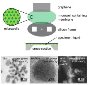
Fig. 1: Structure of the graphene-supported microwell liquid cell architecture and TEM/SEM micrographs of observed processes.
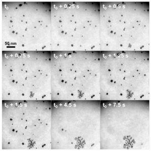
Fig. 2: Image series of gold dendrite growth from HAuCl4-precursor solution.
References
[1] N. de Jonge et al., PNAS 106 (2009) 2159.
[2] M. J. Williamson et al., Nat. Mater. 2 (2003) 532.
[3] H. Zheng et al., Science 324 (2009) 1309.
[4] A. Hutzler et al., in: Proc. EMC Vol. 1 (2016) 209.
[5] A. Hutzler et al., Microsc. Microanal. 22 (2016) 78.